ADA MED SUPPLY LIMITED
Phone:+86 13383897707
Tel:+86-0379-65160607
Email:adaanatomy@adaanatomy.com


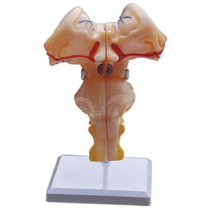
Short answer: The BIX-A1107 is an enlarged brainstem model built from two halves of a sagittal section. It shows the parts of the brainstem — midbrain, pons and medulla — together with the inter-brain (diencephalon) and the cranial nerves, and it carries 67 indications. The model measures 14.5 × 12 × 18 cm and is listed at a reference price of US $61.88, subject to order quantity.
Key takeaways
| Item | Specification on the product page |
|---|---|
| Construction | Two parts of the brainstem sagittal section |
| Structures shown | Midbrain, pons, medulla, inter-brain and cranial nerves |
| Labelling | 67 indications |
| Size | Enlarged — 14.5 × 12 × 18 cm |
| Reference price | US $61.88 (subject to order quantity) |
| Factory certifications | ISO 9001, ISO 14001, ISO 45001 |
The brainstem is where students stop being able to learn by picture. Dozens of named structures sit within a few centimetres of each other, and the clinical consequences of confusing them are severe — a lesion a few millimetres away changes the whole presenting syndrome.
Physically, the region is small and deeply placed, which is exactly why a scaled-up sagittal specimen earns its place on the bench. The comparative evidence supports the format: a meta-analysis of comparative studies found that physical models are effective in anatomy teaching [1], and a later controlled comparison reported that three-dimensional physical models outperformed two-dimensional computer presentations for anatomy learning [2]. For a structure whose whole difficulty is spatial, that distinction is not academic.
There is also a functional reason to pair structure with purpose. The brainstem is not a map of names but the seat of vital automatic function: the pre-Bötzinger complex, a small region of the ventral medulla, was identified as a site that may generate the respiratory rhythm in mammals [3]. A student who has physically separated the medulla from the pons has a far better chance of remembering which functions sit where.
Q1. What is the BIX-A1107?
It is an enlarged brainstem model consisting of two parts of the sagittal section, showing the midbrain, pons, medulla, inter-brain and cranial nerves with 67 indications.
Q2. How large is the model?
It is an enlarged model measuring 14.5 × 12 × 18 cm.
Q3. How many structures are labelled?
Sixty-seven indications are marked on the model.
Q4. Can the model be taken apart?
Yes. It is supplied as two parts of the brainstem sagittal section, so the internal surface can be shown separately from the external one.
Q5. What does it cost?
The page lists a reference price of US $61.88, subject to order quantity. Request a written quotation for the exact volume you need.
Q6. How do I request a quotation or the label list?
Email adaanatomy@adaanatomy.com with the model (BIX-A1107), the quantity you are considering and your destination port; our export team will reply with pricing, lead time and configuration details.
Email adaanatomy@adaanatomy.com for the BIX-A1107 price list and shipping details. If your programme covers the rest of the nervous system, the same brain-model range includes artery-and-nerve brain models and electric brainstem models, so a full neuroanatomy teaching set can be specified in one order.
