ADA MED SUPPLY LIMITED
Phone:+86 13383897707
Tel:+86-0379-65160607
Email:adaanatomy@adaanatomy.com


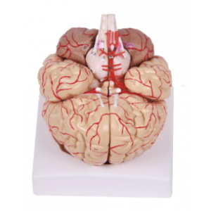
Short answer: The BIX-A1049 is a nine-part anatomical model of the human brain that shows both structure and supply. It separates into the sagittal section of the brain, the cerebral hemisphere, the cerebellum, the brainstem and the cerebral vessels, and it displays the diencephalon, pons, medulla, cranial nerves and cerebrovascular system across 127 individual structures. At natural-large size — 14 × 13 × 16 cm — it fits on a laboratory bench while still being large enough to trace a named artery from origin to territory.
Key takeaways
| Component / region | What students can examine |
|---|---|
| Cerebral hemisphere | Surface topography, lobes, sulci and gyri |
| Sagittal section | Midline structures, corpus callosum, ventricles, diencephalon |
| Cerebellum | Hemispheres and vermis |
| Brainstem, pons and medulla | Continuity from midbrain to medulla, cranial nerve attachment |
| Cerebral vessels | Arterial supply and its relationship to the structures it perfuses |
| Cranial nerves | Course and relation to brainstem and skull base landmarks |
Because the vessels are modelled together with the brain substance, students can answer the question that pure-surface models cannot: which structure is at risk if this vessel is occluded?
Neuroanatomy is the part of gross anatomy where two-dimensional images fail most often, because the structures that matter sit in three planes at once. The evidence supports adding a physical model rather than replacing dissection with more slides.
In a randomised educational study at Boston University School of Medicine, first-year medical students who built and handled 3D colour-coded physical models of the periventricular structures scored significantly higher overall than a control group that re-examined 2D brain cross-sections during laboratory review (t(85) = 2.02, P < 0.05). When the questions were split by the type of thinking they required, only the questions needing 3D visualisation were significantly better in the model group (P = 0.02) — the physical model improved spatial understanding specifically, which is the skill neuroanatomy demands. 84% of the students asked for the activity to be repeated in future laboratories [1].
That finding matches the wider meta-analytic picture. Across 36 studies and 2,226 participants, three-dimensional visualisation technologies produced better factual knowledge (d = 0.30, 95% CI 0.02–0.62) and significantly better spatial knowledge acquisition (d = 0.50, 95% CI 0.20–0.80) than other teaching methods, with higher learner satisfaction [2].
The clinical context makes the spatial skill worth the bench space. Cerebrovascular disease sits inside the world's leading cause of death: an estimated 19.8 million people died from cardiovascular disease in 2022, approximately 32% of all global deaths, and 85% of those deaths were caused by heart attack and stroke [3]. A student who has traced the middle cerebral artery with their hands is better equipped to relate a territory to a deficit at the bedside.
Q1. What is the BIX-A1049?
A nine-part anatomical model of the human brain with artery and nerve, showing the cerebral hemisphere, diencephalon, cerebellum, brainstem, pons, medulla, cranial nerves and cerebrovascular system across 127 structures.
Q2. How big is it?
It is manufactured at natural-large size, 14 × 13 × 16 cm.
Q3. How many parts does it disassemble into?
Nine components, including the sagittal section of the brain, the cerebral hemisphere, the cerebellum, the brainstem and the cerebral vessels.
Q4. Is it suitable for medicine, nursing and high-school biology?
The model is detailed enough for undergraduate neuroanatomy and basic science courses. For an introductory course, ask the supplier to compare it with the simpler 3-part brain models in the same line.
Q5. What is the price?
The product page lists a reference price of US $88.40, subject to order quantity. Email for a formal quotation.
Q6. How do I order, or request a comparison with other brain models?
Email adaanatomy@adaanatomy.com with the model (BIX-A1049), the quantity and your destination; the team will send pricing, lead time and a comparison against the other brain, skull and torso models.
Email adaanatomy@adaanatomy.com for the BIX-A1049 price list and shipping details. The same catalogue covers skull with brain models, whole-body skeletons and sectioned trunks, so a neuroanatomy bench and a general anatomy bench can be specified together.
