ADA MED SUPPLY LIMITED
Phone:+86 13383897707
Tel:+86-0379-65160607
Email:adaanatomy@adaanatomy.com


Article tag: Female Torso Vertical Slice Anatomical Model BIX-A3005 A3005
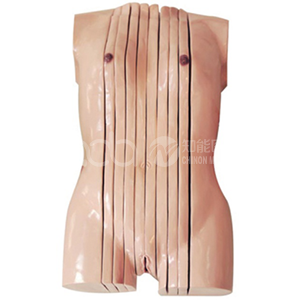
Model | BIX-A3005 — Female Torso Vertical Slice Anatomical Model |
Summary | Female torso vertical slice model in 10 dissectible sagittal parts, 42 x 20 x 84.5 cm, showing internal organs in section for anatomy teaching. |
Construction | A total of 10 parts (manufacturer's specification), dissectible |
Dimensions | 42 x 20 x 84.5 cm |
Section Plane | Vertical slice — a sagittal section through the female trunk, showing internal organs in section |
Certification / Price | ISO & CE (as stated); US $1,267.08 |
Educational-use note: anatomy teaching model — educational equipment, not a medical device.
Before ordering: the page gives two specification lines only (10 parts; 42 x 20 x 84.5 cm), there is no specification PDF, and the price appears on the category listing but not in the product page body. Request the datasheet and part list: adaanatomy@adaanatomy.com.
The A3005 is a vertical slice of the female trunk — 10 dissectible parts, 84.5 cm tall, 42 cm wide, 20 cm deep. The page spec is two lines; the meta description adds that it shows internal organs in sagittal section for teaching the structure of the female body.
It matters most as one of three planes, because the same torso is offered in all three radiological planes — and that is the real buying decision:
Plane | Model | Parts | Price |
Sagittal (vertical slice) | A3005 female / A3004 male | 10 | US |
Transverse (10 mm serial) | A3007 male / A3008 female | — | US |
Coronal (frontal) | A3009 | 8 | US $1,478.06 |
The planes are not interchangeable — the one you choose decides which clinical skill the model can build.
It also sits far above the conventional torso range — A1043 (85 cm, 40 parts, US  185.64), ADA-210 (US $283.76). At roughly 4.5× the most expensive of those, the premium buys the section plane, not more parts.
Clinical image interpretation is a 3D-to-2D translation. One paper states the problem directly: it means identifying three-dimensional anatomical features in two-dimensional cross-sectional CT and MRI images in axial, sagittal and coronal planes, requiring the student to reconcile known 3D structures with previously unknown 2D visual information — a cognitive transition the authors say should be taught by combining 3D and 2D approaches.
That is the argument for a vertical slice torso. A conventional torso opens at the front like a book: good for gross anatomy, useless for reading a CT slice. A model itself cut in a plane matches the picture on screen — same plane, same relationships, same loss of the third dimension.
Sagittal slices have been tested as an adjunct. A randomised educational trial compared sagittal plastinated slices (SPS) against conventional resources for foot and ankle anatomy, measured by learners' ability to correctly identify structures on sagittal MRI images. Scores rose a mean of 1.2 against 0.7 for controls, out of 14 — not statistically significant (p = 0.41). Learners reported SPS was most useful after a session using conventional resources.
Read that as a usage instruction: a sagittal-sectional model is an adjunct applied after the basics — after the open-front torso, not instead of it.
Against dissection and 3D software. A study of 313 medical students taught upper-limb anatomy through dissection (n = 80), prosections (n = 77), plastic models (n = 84) and 3D software (n = 72), examined with 100 multiple-choice and tag questions. Dissection and the 3D group outperformed the prosection and the plastic models group in total and multiple-choice questions, and the 3D group's tag-question performance was significantly higher than the other three.
Against non-cadaveric teaching generally. A study of 120 first-year medical students grouped by practical exposure — cadaver dissection (n = 69) versus non-cadaveric methods only (n = 51) — found the cadaver group scored higher on multiple-choice questions (69.90% vs 55.79%) and essays (61.16% vs 52.92%). Cadaver dissection faces shortages, costs and curriculum changes, and comparative evidence "remains mixed."
What this means for a buyer. A plastic model is not a proven substitute for dissection — and neither study tested a sectional model. The defensible claim is narrower: the A3005 is the only physical object in the room matching a CT slice plane.
Patient and consent education is where sectional models show measurable effects. In a study of seven patients undergoing percutaneous nephrolithotomy, personalised 3D-printed kidney models were used during informed consent. Before-and-after questionnaires were significant for understanding of kidney anatomy, stone size, the procedure and satisfaction (p = 0.046, 0.025, 0.046 and 0.046), and five of seven patients (71.4%) called the model very useful. None of the patients answered that the cost was appropriate — the most honest sentence in this literature, and it applies to a US $1,267 torso.
A second study of seven patients shown life-size patient-specific models reported gains in kidney anatomy of 50% (p = 0.026) and the planned surgical procedure of 44.6% (p = 0.026).
Both are pilots of seven patients — a coherent direction, not an established effect size: enough to trial a sectional model, not to justify a budget line alone.
Where it sits. The A3005 is the female sagittal member: A3004 (male sagittal, US 1,458.60 / US 1,478.06). Below it: A1043 (US 81.04), A1044 / A1045 (US $64.82).
Choose the plane from the skill: sagittal (A3005 / A3004) for midline relationships; transverse (A3007 / A3008) for serial axial reading, the plane most reporting uses daily; coronal (A3009) for frontal-plane work with fewer, larger parts.
Buy the A3005 when a programme teaches gross anatomy adequately already and needs to add cross-sectional reasoning before students meet real CT and MRI.
Checklist (cross-sectional drill)
● Oriented on the model's plane before any image is shown
● One structure traced from the physical slice to the matching CT or MRI slice, same plane
● Midline structures named and distinguished from overlying organ positions
● The model used after
conventional open-front teaching, not as its replacement
Q1: What is the BIX-A3005? A: A female torso vertical slice anatomical model — a sagittal-section trunk in 10 dissectible parts, 42 x 20 x 84.5 cm.
Q2: How does it differ from a conventional anatomy torso? A: A conventional torso opens at the front and shows organs in normal positions. The A3005 is cut in a vertical plane, matching a sagittal CT or MRI slice.
Q3: Which plane should I buy? A: Sagittal (A3005 / A3004) for midline relationships; transverse (A3007 / A3008) for serial axial reading; coronal (A3009) for frontal-plane work.
Q4: What is the price? A: US $1,267.08. Email adaanatomy@adaanatomy.com for quotation, part list, bulk pricing and the datasheet.
Q5: Does the evidence support models over dissection? A: Not uniformly. A study of 313 students found dissection and 3D software outperformed plastic models, so a sectional model is best used as an adjunct after conventional teaching.
Q6: What documentation is available? A: ISO & CE as stated. The page publishes only two specification lines and no PDF — request the datasheet: adaanatomy@adaanatomy.com.
Comparing Anatomy Teaching Modalities: Dissection, Prosections, Models (2021)
Cadaveric vs Non-Cadaveric Anatomy Teaching in Nigerian Students (2026)
Sagittal Plastinates for Foot and Ankle Anatomy: Randomized Trial (2019)
3D-2D Transitions in Anatomy Learning (2020)
3D-Printed Kidney Model for Patient Understanding of PCNL (2022)
Personalized 3D-Printed Kidney Model for Patient Education (2016)
