ADA MED SUPPLY LIMITED
Phone:+86 13383897707
Tel:+86-0379-65160607
Email:adaanatomy@adaanatomy.com


Article tag: Comprehensive Skills Training Model for Childbirth BIX-F50 F50
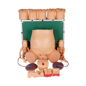
Model | BIX-F50 — Comprehensive Skills Training Model for Childbirth (Obstetric Delivery Trainer) |
Summary | Childbirth training model: pregnant lower body, two fetuses, cord and placenta; six cervical dilatation stages, soft perineum and transparent abdominal wall. |
Configuration | Simulated pregnant lower body; two fetal models; cord and placenta models |
Cervical Checking Model | Six stages — not dilated, not effaced, station −5 · 2 cm, 50% effaced, −4 · 4 cm, −3 · 5 cm, 0 · 7 cm, +2 · 10 cm, +5 |
Fetal Model | Soft skin; discernible fontanelle for practising aspiration |
Delivery Model | Soft, highly flexible perineum; also used for nursing skills |
| Abdominal Wall | Transparent wall plus simulation skin, so the delivery demonstration and the head's relationship to the ischial spine plane can be observed | | Stated Purpose | Obstetric basic training: prenatal examination, midwifery and delivery skills | | Certification / Price | ISO 9001 / 14001 / 45001 & CE (page-stated); US $883.80 |
Educational-use note: educational equipment, not a medical device for patient use. Request the spec sheet by email for spare fetuses, cords and placentas.
The F50 is three teaching devices sharing one pelvic body.
A staged cervix. The cervical checking model is built as six discrete stages, each pairing a dilatation with an effacement state and a station against the ischial spine plane — from no dilatation at −5 to 10 cm at +5 — a progression an instructor can set by hand.
A fetal unit with annexes. A fetal model with soft skin and a discernible fontanelle supports aspiration practice, and the set includes cord and placenta models.
A delivery pelvis with a window. The perineum is deliberately soft and highly flexible, and the abdomen combines a transparent wall with simulation skin so the delivery demonstration — and the head's relationship to the ischial spine plane — can be seen rather than inferred.
Stated purpose: obstetric basic technical training — prenatal examination, midwifery and delivery skills — at US $883.80, ISO 9001 / 14001 / 45001 and CE.
The strongest argument comes from a study using almost the same apparatus: a model fetal head inside a mannequin's pelvis, assessed by 20 midwives and 20 doctors at five randomly set stations between −2 and +7 cm. Mean error (SD) ranged from 0.3 (1.3) to 4.3 (1.1) cm for digital examination against 0.1 (1.2) to 2.6 (1.6) cm for a reference device; inaccuracies increased as the head descended, and digitally the true value fell outside one standard deviation for stations beyond +1 cm (Awan et al., 2009).
Current literature states that difficult-labour decisions "rest almost entirely on 3 pieces of information obtained by digital vaginal examination: the position of the fetal occiput, the attitude of the fetal head, and the station of the leading bony point", with the examination "least reliable when the clinical stakes are highest" because caput, moulding and a high presenting part obscure the sutures and fontanelles it depends on (Mappa & Rizzo, 2026).
Inaccurate assessment of position and station "might increase the risk for difficult or failed assisted vaginal delivery" (Chan & Lau, 2021); the soft perineum is where controlled delivery is first practised, and the transparent wall lets a group watch descent.
An honest review has to draw the boundary clearly.
It cannot remove inter-observer variability. Agreement between ultrasound and vaginal examination across position, station and progression was reached in only 27 of 47 patients (57%), with caput succedaneum present in 45% of the non-correlated cases (Borovska et al., 2026). A clean silicone cervix will not reproduce the caput and moulding behind that disagreement.
It cannot make the clinical call objective. Digital estimation accuracy was 34% in a study of 62 term women, rising to 74.19% only with a ±1 cm tolerance (Messina et al., 2021). And its six stages are a simplification: dilatation is continuous, and labour progress assessment has "remained largely unchanged for at least a century" while remaining "inconsistently reproducible" (Usman et al., 2023).
No training model changes outcomes by itself. Even intrapartum ultrasound, which outperforms digital examination statistically, has not yet demonstrated a difference in obstetric and neonatal morbidity and remains an adjunct, not a replacement (Mappa & Rizzo, 2026).
Dimension | Rating | Basis |
Cervical dilatation realism | Good | Six graded stages, effacement and station paired |
Fetal station training | Very good | Ischial spine plane modelled; station −5 to +5 |
Delivery demonstration | Very good | Transparent wall lets descent be observed |
Caput and moulding | Limited | Not represented; the main real confounder |
Value at US $883.80 | Good | Replaces separate pelvis, cervix and fetal units |
Model | Scope | Best for |
F50 | Staged cervix + two fetuses + transparent-wall pelvis | Assessing dilatation and station |
F8 | Standard delivery programme, true-to-size pelvis | Core normal-birth sequence |
F8A | Advanced dystocia training | Shoulder dystocia |
F54 | Abdominal palpation plus delivery mechanism | Antenatal palpation |
F9 | Advanced cervical change model | Cervical change over time |
Buying logic: buy the F50 where students must learn to assess dilatation and station, not merely to deliver; add F8A for dystocia.
Station | Time | Activity |
A. Baseline blind round | 15 min | Set a station; student estimates it before teaching |
B. Stage map and landmarks | 30 min | All six stages; fontanelle and ischial spines related to the station scale |
C. Delivery with a window | 25 min | Deliver with the transparent wall in place; narrate descent |
D. Re-test | 15 min | Repeat the blind round; compare error with the baseline |
Assessment checklist
● All six stages identified with dilatation, effacement and station together
● Station named relative to the ischial spine plane
● Fontanelle located and aspiration technique demonstrated
● Blind-round error reduced against the student's own baseline
Item | Frequency | Notes |
Pelvis, perineum and abdominal window | Each session | Mild disinfectant; no abrasives on the window |
Fetal models, cord and placenta | Each session | Check for tears; store uncompressed |
Cervical stage inserts | Monthly | Confirm each stage seats correctly |
Q1: What exactly does the BIX-F50 include? A: A simulated pregnant lower body, two fetal models, cord and placenta models, a six-stage cervical checking model, and a delivery model with a soft perineum and transparent abdominal wall.
Q2: Which dilatation stages does the cervix show? A: Six stages from no dilatation at station −5 to 10 cm at +5 (see the table above).
Q3: What is the transparent abdominal wall for? A: It lets an instructor or a group observe the delivery demonstration and the head's relationship to the ischial spine plane directly.
Q4: Can students practise fetal aspiration? A: Yes — the fetal model has soft skin and a discernible fontanelle.
Q5: What is the price and MOQ? A: Listed at US $883.80; MOQ 1 unit — email adaanatomy@adaanatomy.com..
Q6: What certifications and shipping terms apply? A: ISO 9001, ISO 14001, ISO 45001 & CE as stated on the page; air freight 7–10 days, sea freight 30–45 days. Details: adaanatomy@adaanatomy.com..
Accuracy of Digital Station Assessment in a Pelvic Model (Awan et al., 2009)
Digital Examination Versus Intrapartum Ultrasound (Borovska et al., 2026)
Accuracy of Vaginal Examination for Head Station (Messina et al., 2021)
The Sonopartogram Review (Usman et al., 2023)
Intrapartum Sonography for Difficult Labour (Mappa & Rizzo, 2026)
Ultrasound and the Mode of Delivery Decision (Chan & Lau, 2021)
