ADA MED SUPPLY LIMITED
Phone:+86 13383897707
Tel:+86-0379-65160607
Email:adaanatomy@adaanatomy.com


Article tag: Life-Size Adult Female Pelvic Skeleton Model BIX-A1036 A1036
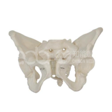
Model | BIX-A1036 — Life-Size Adult Female Pelvic Skeleton Model |
Summary | Life-size female pelvic skeleton model: low wide pelvis, barrel-shaped cavity, pubic arch 90-100 degrees. For obstetric anatomy teaching. (137 chars) |
Material | PVC |
Scale | Natural large (life-size) |
Female Features | Low, wide pelvis · barrel-shaped pelvic cavity · wide, short pubic symphysis · pubic arch 90–100° |
Related | A1035 (male adult pelvis, for sex-difference comparison) · A1038 (female pelvis with fetal skull) |
Price | On request |
Audience | Medical schools, midwifery/OB-GYN programs, osteopathic and PT education, hospital training departments |
Educational-use note: anatomical teaching model — educational equipment, not a medical device or pharmaceutical product, and contains no medicinal ingredients (per manufacturer's page notice). One line of the manufacturer's web text (referring to a "tibia" shape) appears to be a typographical error; the classic female-pelvis features listed above follow the rest of the official description and standard anatomy, and should be confirmed against the specification sheet.
The pelvis is arguably the most challenging region in anatomy education: it is three-dimensionally complex, contains reproductive, urinary, and digestive systems in a compact bony frame, and its spatial relationships are hard to grasp from 2D atlases or dissections alone. Novice students struggle with the pelvis and perineum "due to the complex arrangement and limited visibility of structures in these areas" (interactive pelvic model study, 2017).
For clinical programs the stakes are high:
Obstetrics:
● pelvic shape and capacity directly inform delivery-mechanism teaching and the assessment of labor progression.
Gynecology & urology:
● bony landmarks anchor surgical approaches and catheterization teaching.
Orthopedics & physical therapy:
● the pelvic ring and its sex differences underpin gait, posture, and pelvic-floor education.
Yet pelvic anatomy is a documented knowledge deficit in OB/GYN residents — traditional teaching methods have obstacles and are often costly (clay-pelvis study, 2022). A dedicated life-size female pelvis model is therefore not an accessory; it is the anchor object of an entire teaching station.
Evidence | Finding | Implication |
Clay-pelvis study (2022, PubMed 35849463) | OB/GYN residents building a model on a bony pelvis: 66.7% improved knowledge (up to 30%); self-assessed confidence rose significantly (P=.010–.019); high satisfaction | A real bony pelvis + hands-on layering is the foundation residents respond to |
Interactive pelvic model study (2017, PubMed 28885415) | Faculty and learners positively evaluated a reusable pelvic model for teaching 3D anatomic relationships | Physical 3D models close the 2D-atlas gap in pelvic teaching |
Fleming et al., 2020 (J Am Coll Radiol) | 3D-printed/physical models significantly raised anatomy exam performance in medical students (P<0.0001) | Physical models outperform 2D teaching across anatomy |
Cook et al., 2011 (JAMA) | Technology-enhanced simulation yields large learning gains (effect sizes 1.09–1.20; 35,226 learners) | Hands-on model-based teaching is among the most effective methods |
The teaching logic is simple: a life-size female pelvis lets learners see, touch, and relate — the entry shape, the cavity, the arch, the symphysis — instead of memorizing drawings.
Feature (official spec) | What It Demonstrates |
Pelvis full shape: low and wide | The classic gynecoid (female) overall proportion — the entry width-to-height relationship that differs from the male pelvis |
Pelvic cavity: barrel-shaped | The roomier female cavity shape relevant to obstetric teaching |
Pubic arch: 90–100° | The wide female pubic arch (vs the narrow male arch <90°) |
Pubic symphysis: wide and short | Female symphysis morphology vs the taller male symphysis |
Life-size scale | Anatomically correct proportions for palpation and landmark drills |
Model | Role |
A1035 | Male adult pelvis — side-by-side sex-difference teaching |
A1036 | Female adult pelvis — obstetric morphology anchor |
A1038 | Female pelvis with fetal skull — engagement and mechanism-of-labor visualization |
Programs teaching obstetric anatomy typically run the pair: A1035 vs A1036 for sex differences, then A1038 (or F9) to connect pelvic shape to labor mechanics.
1. Place A1036 and A1035 side by side.
2. Compare entry shape, arch angle, cavity depth, symphysis height.
3. Relate each difference to its obstetric or functional meaning.
1. Palpate and name key landmarks: pubic symphysis, ischial spines, sacral promontory, iliac crest.
2. Associate each landmark with clinical use (e.g., ischial spines — station assessment in labor).
1. Use A1038 (fetal skull) or diagrams to show engagement and descent through the female cavity.
2. Connect pelvic morphology to delivery-mechanism teaching (rotation, extension).
Criterion | Standard |
Landmark identification | Correct naming and palpation of major pelvic landmarks |
Sex differences | Correctly list ≥5 male–female pelvic differences |
Obstetric relevance | Correctly explain arch/cavity significance for labor |
Spatial relations | Correctly describe entry→cavity→outlet axis |
Item | Frequency | Notes |
PVC surface | After each use | Wipe clean; PVC is washable |
Storage | Always | Dust-free, dry; avoid prolonged direct sunlight |
Handling | Always | PVC is durable; drop from height not advised |
Q1: What is the difference between A1036 and A1035? A: A1036 is the female adult pelvis (low, wide, barrel-shaped cavity, 90–100° pubic arch); A1035 is the male adult pelvis. The pair is used side by side to teach sex differences in pelvic morphology — a core anatomy objective.
Q2: How does this model connect to obstetric teaching? A: Pelvic shape and capacity are the anatomical frame for delivery-mechanism education. Teaching it alongside A1038 (female pelvis with fetal skull) or labor models lets students visualize how fetal descent relates to the female pelvis.
Q3: Who should use this model? A: Medical schools, midwifery and OB-GYN programs, osteopathic/PT programs, and hospital training departments. It is the anchor object for pelvic anatomy stations and OB orientation.
Q4: Does hands-on pelvic teaching actually improve learning? A: Yes — OB/GYN residents who built on a real bony pelvis improved knowledge (66.7% gained up to 30%) and significantly raised confidence (P=.010–.019) (clay-pelvis study, 2022), and physical 3D models significantly raise anatomy exam performance (Fleming et al., 2020).
Q5: What material is it made of? A: PVC — durable, washable, and suitable for repeated handling in classroom use.
Q6: What is the MOQ and delivery time? A: MOQ is 1 unit. Air freight: 7–10 business days; sea freight: 30–45 days. Email adaanatomy@adaanatomy.com for the specification sheet and quote.
Effectiveness of a Clay Pelvic Model: A Hands-on Approach to Understanding Pelvic Floor Anatomy (2022)
Interactive Pelvic Anatomy Model: A Tool for Teaching Basic Pelvic Anatomy (2017)
Effectiveness of Three-Dimensionally Printed Models in Anatomy Education for Medical Students and Resident Physicians: Systematic Review and Meta-Analysis — Fleming et al. (2020), J Am Coll Radiol 17(10):1220–1229
Technology-Enhanced Simulation for Health Professions Education: A Systematic Review and Meta-analysis — Cook et al. (2011), JAMA 306(9):978–988
