ADA MED SUPPLY LIMITED
Phone:+86 13383897707
Tel:+86-0379-65160607
Email:adaanatomy@adaanatomy.com


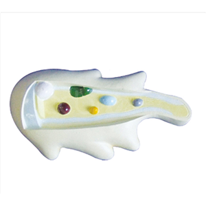
Short answer: BIX-C0001 is an enlarged model of Amoeba proteus built to show one cell in enough size and colour separation that a class can see its parts at once — cell membrane, cytoplasm, contractile vacuole and pseudopodia. It is a structural teaching model, not a specimen: the parts are colour-separated for instruction, and the material is plastic at a published size of 9.44 × 5.51 inches.
Key takeaways
| Published part | What the lesson is |
|---|---|
| Cell membrane | The boundary of a single-celled organism — where exchange with the environment happens. |
| Cytoplasm | The interior in which everything else is suspended; the reason the cell is treated as a system rather than a shape. |
| Contractile vacuole | Water regulation. This is the structure that turns "a cell is a bag" into "a cell maintains conditions". |
| Pseudopodia | Locomotion and feeding — the visible link between structure and behaviour. |
| Item | Published value |
|---|---|
| Product model | BIX-C0001 |
| Category | Biology Series → Animal Models |
| Size | 9.44 × 5.51 inches |
| Material | Plastic |
| Finish | Environmentally friendly paint, colour-separated by part |
| Price | Inquiry for best offer — subject to order quantity |
| Factory certification shown on page | ISO 9001, ISO 14001, ISO 45001 |
The value of a protozoan model is that it keeps one cell as the unit of study while making it large enough to inspect. That matters because the alternative — a microscope image — is a two-dimensional projection, and the literature on anatomy learning has been fairly consistent that a physical object carries spatial information that a flat presentation does not. A randomised comparison found three-dimensional physical models outperformed two-dimensional computer presentations for learning anatomy [1], and the same conclusion has been supported at review level for physical models in anatomy teaching generally [2].
The same species also appears in the research literature on cell behaviour, which is useful when a student asks whether this is a "textbook animal". Research on Amoeba proteus has examined how anticipation of periodic events influences cell motility [3]. In other words, the structures this model colour-codes are the structures still being measured in current work — the model is a scale model of an active research subject, not a textbook cartoon.
| Not stated on the page | Why you need it |
|---|---|
| Magnification factor | The page gives size, not enlargement ratio; confirm if the ratio matters for your curriculum. |
| Base or stand | Determines whether it can be displayed upright or must be laid flat. |
| Packaging dimensions and gross weight | Needed to calculate landed cost for a multi-unit biology order. |
| Price at your order quantity and lead time | Pricing is by enquiry and quantity-dependent. |
Send these questions to adaanatomy@adaanatomy.com with the model number BIX-C0001 and your quantity, and we will answer each line and attach the current supply data.
Q1. What is BIX-C0001?
It is an enlarged model of the amoeba Amoeba proteus, showing the cell membrane, cytoplasm, contractile vacuole and pseudopodia, with the parts separated by colour.
Q2. What size is it?
The published size is 9.44 × 5.51 inches, in plastic.
Q3. Is it a specimen or a model?
It is a teaching model. The structures are colour-separated to make them readable in class, which a real specimen would not allow.
Q4. What can it teach that a slide image cannot?
Three-dimensional structure and the relationship between parts in space; the model can also be handled and passed round a class.
Q5. Is the price published?
No. The page states "Inquiry for Best Offer", subject to order quantity.
Q6. How do I get a quotation?
Email adaanatomy@adaanatomy.com with the model (BIX-C0001), your quantity and the other biology models you are considering, so they can be quoted together.
Write to adaanatomy@adaanatomy.com for the BIX-C0001 specification sheet and a quotation. If you are building a biology teaching set, ask about the Paramecium model and the plant-cell models in the same enquiry so the whole set ships together and the carton data can be planned once.
