ADA MED SUPPLY LIMITED
Phone:+86 13383897707
Tel:+86-0379-65160607
Email:sophia@adahealthy.com


Article tag: Leg venipuncture model venipuncture model
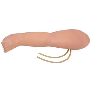
Venipunction in the legs is a common but challenging procedure in medical practice, especially for elderly patients, obese patients, or patients with poor venous conditions. However, as medical technology continues to advance, the leg venipunction model has emerged to provide healthcare workers with an efficient and safe training platform that allows them to easily tackle this challenge.
First of all, medical staff can perform repeated puncture exercises on the model, familiarize themselves with the characteristics of veins in different parts and under different conditions, and master the correct puncture Angle and depth. This simulated training not only improved the piercing skills of the medical staff, but also increased their confidence, making them more comfortable in the face of real patients.

Secondly, it has a variety of functions to meet the training needs of different levels. For example, some advanced models can simulate the difficulty of puncture under different conditions, such as venous sclerosis, edema, etc., to help medical staff master ways to deal with these complex situations. In addition, the model can also provide immediate feedback, such as whether the puncture is successful, whether there are complications, etc., to help medical staff timely adjust the manipulation and improve the success rate of puncture.
Most importantly, the use of the leg venipunctures model greatly reduces the pain and risk for patients. In the traditional teaching mode, medical staff often need to practice on real patients, which not only increases the pain and discomfort of patients, but also may lead to complications due to improper operation. With it, medical staff can fully practice in a simulated environment, reducing errors and uncertainties in actual operation, thus ensuring the safety and comfort of patients.
To sum up, venous puncture models of the legs are an indispensable and important tool in medical training. It helps healthcare professionals easily navigate the challenges of leg venipentesis by simulating real-world environments, providing immediate feedback, and reducing patient risk. With the continuous development of medical technology, we have reason to believe that leg venipuncture models will play an even more important role in medical training in the future.
