ADA MED SUPPLY LIMITED
Phone:+86 13383897707
Tel:+86-0379-65160607
Email:sophia@adahealthy.com


Article tag: Full-featured neonatal simulator Neonatal model
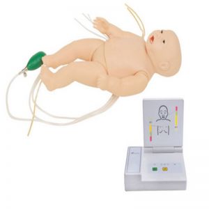
In the wave of medical technology, the fully functional neonatal advanced simulator is like a giant ship, leading us into a new era of medical training. This simulator not only integrates cutting-edge simulation technology, but also achieves the ultimate in details, providing a nearly realistic training environment for medical staff.

Imagine that with the help of a simulator, medical staff can simulate various physiological states and pathological changes of newborn babies. From weak breathing to strong heartbeat, from rosy skin color to pale appearance, the simulator can present it one by one. Such a training environment undoubtedly provides a valuable practice opportunity for medical staff, allowing them to fully practice and become familiar with various neonatal care skills in a safe environment.
What’s more worth mentioning is that the full-featured advanced newborn simulator also has a high degree of interactivity and intelligent evaluation functions. Medical staff can freely set the simulator's condition and response as needed to conduct targeted training. At the same time, the simulator can also record the operation process of medical staff in real time and score according to preset standards, so that medical staff can understand their own shortcomings in time and continuously improve their skill level.
The emergence of this simulator not only improves the training effect of medical staff, but also provides a solid guarantee for the life safety of patients. In the future medical field, fully functional neonatal advanced simulators will play an increasingly important role and become an indispensable training partner for medical staff.
In this era full of changes, the fully functional neonatal advanced simulator leads the new trend of medical training with its unique advantages. We believe that with its help, medical staff will be able to better master neonatal care skills and protect the lives of patients.
